Radical oxygen species formation in metabolic enzymes

The mitochondria in our cells are responsible for the conversion of energy from nutrients into ATP, the native fuel of the organism. However, occasional stray chemical reactions in the mitochondria lead to production of a small amount of reactive oxygen species (ROS), such as superoxide, which are harmful to cells and related to aging [Massaad et al. Aging 1:758. 2009, Brand. Exp. Gerontol 45:466. 2010]. As the metabolic energy conversion processes often involve a series of redox reactions, or electron transfers, and the enzymes involved therefore contain redox active cofactors or prosthetic groups, it is conceivable that occasional reactions with e.g. molecular oxygen could lead to small amounts of ROS formation as an unwanted byproduct. Computational modeling of processes, including binding and unbinding of oxygen molecules and electron transfer to O2, have been carried out for two important metabolic enzymes to shed light on possible ROS formation mechanisms in the metabolic pathways.

Cytochrome bc1 complex

The membrane-embedded cytochrome bc1 complex, also known as complex III in the electron transport chain is one of the central enzymes in the energy conversion machinery of mitochondria and is believed to produce small yields of superoxide through side reactions with molecular oxygen. The mechanism of such superoxide production in the bc1 complex is, however, still unclear.

The bc1 complex serves to transport protons across the membrane through a series of redox reactions with substrate molecules in the membrane to maintain a cross-membrane potential, which is used to power ATP synthesis. The net reactions and transport processes during the enzyme's complex reaction cycle, the Q-cycle, are depicted in Fig. 1. At intermediate stages, substrate quinol molecules become radical semiquinones, which conceivably could further lead to production of other radical species such as superoxide.

The Q-cycle of the bc1 complex
Figure 1. During the reaction cycle of the bc1 complex, the Q-cycle, two protons are absorbed from the negative side of the membrane, while four protons are released to the positive side. This process is fueled by redox reactions with substrate quinol (QH2) and quinone (Q) molecules from the membrane: two QH2 are oxidized to Q at the Qo binding site of the bc1 complex, while one Q molecule is reduced to QH2 at the negative side. The process is coupled to a series of internal electron transfers in the bc1 complex.  The reaction cycle described here applies to each of the two identical functional monomers making up the dimeric bc1 complex.


The proton transfers in the bc1 complex are coupled to a series of internal electron transfers as illustrated in Fig. 2 (see also Proton-coupled electron transfer in the cytochrome bc1 complex), involving several iron-containing prosthetic groups: Three heme groups and an iron-sulfur cluster (Fe2S2). The substrate QH2 at the Qo-site first transfers an electron the Fe2S2-cluster and briefly becomes a radical semiquinone before it is fully oxidized to Q through the second electron transfer to heme bL. An oxygen molecule, if present at the Qo-site, could conceivably "steal" the excess electron from the semiquinone and escape the bc1 complex as a radical superoxide, possibly leading to cell damage.

Electron transfers in the bc1 complex
Figure 2. Many internal electron transfers are involved in the Q-cycle of the bc1 complex. After QH2 binds to the Qo-site, it first transfers an electron to the Fe2S2 cluster, leaving it briefly as a radical semiquinone intermediate before the second electron transfer to heme bL occurs. At this stage, there is a risk of stray reactions with molecular oxygen leading to formation of superoxide.


Molecular dynamics simulations show that oxygen molecules are indeed able to migrate into the bc1 complex and bind at the Qo-site, where it can stay bound for tens of nanoseconds at a time, while a semiquinone substrate is present (see Fig. 3) [Husen & Solov'yov. JACS 138:12150. 2016]. This makes the Qo-site a viable candidate as a center of superoxide production.

Oxygen molecules diffuse into the bc1 complex and migrate to the Qo-site
Figure 3: Oxygen molecules diffuse into the bc1 complex from the membrane and are able to migrate into the Qo-site. The red surface near the semiqinone molecule and the Fe2S2 cluster in the figure to the right indicates a region, where an O2 molecule occasionally binds and stays for up to tens of nanoseconds.


A follow-up study found the precise O2 binding modes and statistics depended strongly on mutations of a tyrosine amino acid (Y302) at the Qo-site [Husen & Solov'yov. J. Phys. Chem. B 121: 3308. 2016]. Fig. 4 shows different observed O2 binding sites as the tyrosine is replaced by alanine (A302), cysteine (C302) or serine (S302).

Different binding sites for oxygen molecules at the Qo-site of the cytochrome bc1 complex, as the tyrosine Y302 is mutated to alanine, cysteine or serine
Figure 4: Different binding sites for oxygen molecules at the Qo-site of the cytochrome bc1 complex, as the tyrosine Y302 is mutated to alanine, cysteine or serine


Further quantum chemical calculations on snapshots of the Qo-site with bound O2 and semiquinone reveal that several charge transfer mechanisms leading to superoxide are possible [Salo et al. J. Phys. Chem. B 121:1771. 2016]: essentially, the excess electron transferred to the oxygen molecule can either come from the Fe2S2 cluster or from the semiquinone as illustrated in Fig. 5.

Superoxide produced by electron transfer from FeB atom or semiquinone
Figure 5: Superoxide is produced at the Qo-site by electron transfer to an O2 molecule bound there. The figure shows the spin density of unpaired electrons after two possible electron transfer mechanisms. A: The electron is transferred from the FeB atom of the Fe2S2 cluster. B: The electron is transferred from the semiquinone, which is thus oxidized to the non-radical quinone.


The ETF enzyme

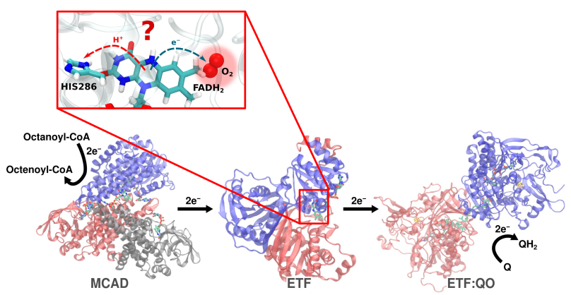
The ETF enzyme, which consists of two protein subunits (alpha and beta) and the cofactors flavin adenine dinucleotide (FAD/ FADH2) and adenosine monophosphate (AMP) facilitates elctron transport as part of the fatty acid metabolism in mitochondria [Rodrigues et al. Free Radical Biol. Med. 53, 12–19 (2012)]. The redox active nature of the protein, however, also makes it a potential source of reactive oxygen species as a byproduct.

Atomistic molecular dynamics simulations were employed [Husen et al. J. Chem. Inf. Model 59, 4868–4879 (2019)] to study the dynamics of oxygen molecules in the ETF enzyme and possible O2 binding modes near the FADH2 cofactor, which could serve as electron donor in a superoxide production mechanism.


The ETF enzyme
Figure 6: The ETF enzyme is part of the fatty acid metabolic pathway in mitochondria. The redox active FADH2 cofactor could occasionally react with a nearby oxygen molecule leading to release of radical oxygen species.


In particular, extensive molecular dynamics simulations of the ETF enzyme in solution with a concentration of oxygen molecules were carried out to identify O2 binding sites and characterize binding modes. Five binding sites were identified near the enzyme’s FADH2 cofactor and were used for further modeling. Site 5 is particularly interesting for superoxide production, as the positively charged β-ARG12 stabilizes the superoxide anion. O2 binding was further characterized through 100 replicate binding event simulations for each of the 5 sites. The characteristic O2 binding time are in the range of 0.5-3 ns, but a subpopulation of simulated events at site 5 showed significantly longer binding.


O2 binding sites in the ETF enzyme
Figure 7: Five O2 binding sites near the FADH2 cofactor of the ETF enzyme were identified. The postively charged (blue) β-ARG12 at site 5 may stabilize binding of superoxide, if a reaction between O2 and FADH2 takes place.


Electron transfer from FADH2 to O2 would initially lead to formation of a radical pair, FADH − O2•− [Imlay, J.A. Nat. Rev. Microbiol 11, 443–454 (2013)]. Interchange between the singlet and triplet states of the radical pair, which could be influenced by external magnetic fields or RF radiation, then decides the ultimate release of reactive oxygen species.


Reaction scheme of radical oxygen species formation

Figure 8: Radical pair dynamics may determine the fate of released reactive oxygen species: depending on the spin state of the radical pair formed through the initial reaction between FADH2 and O2, either H2O2 or superoxide may ultimately be released.